E-Mail: sales@microapproachmed.com
Phone: +86-17722685792
- All
- Product Name
- Product Keyword
- Product Model
- Product Summary
- Product Description
- Multi Field Search

Views: 0 Author: Site Editor Publish Time: 2026-10-27 Origin: Site


In flexible ureteroscopy, the margin between efficient calculus extraction and procedural complication hinges on the precise dimensional tolerances of your chosen extraction instrument. Selecting an incompatible stone retrieval basket compromises working-channel fluid dynamics. It restricts scope deflection in the lower pole and increases the risk of urothelial trauma or entrapment. These devices often double as tools for retrieving migrated stents or foreign bodies, demanding high versatility from your equipment fleet. This guide deconstructs the technical specifications you need to evaluate. We cover wire geometry, nitinol composition, and exact French sizing. You will get a framework for evaluating which configurations align with specific clinical requirements and your existing endoscopes.
Working-Channel Ratios Dictate Performance: Basket sheath sizes (typically 1.5F to 1.9F) must be evaluated against the endoscope’s working channel to ensure adequate irrigation flow and visibility during extraction.
Wire Geometry Drives Capture Mechanics: Flat-wire designs offer superior radial dilation and sweeping capabilities, while round-wire configurations utilize gradual opening mechanics to minimize tissue trauma during complex calyceal manipulation.
Tipless Designs Dominate Lower Pole Access: No-tip (tipless) nitinol baskets allow for closer proximity to the urothelium, enabling the capture of mucosal-embedded fragments while maintaining full scope deflection.
Access Strategy is Interdependent: Optimal basket performance requires concurrent evaluation of ureteral access sheath selection to ensure seamless multi-pass efficiency and controlled intrarenal pressures.
The operational efficiency of flexible ureteroscopy relies heavily on maintaining clear visual fields through continuous irrigation. Introducing any instrument into the endoscope's working channel immediately alters fluid dynamics. There is a strict inverse relationship between the cross-sectional area of the sheath and the volumetric flow rate of the irrigation fluid. When a large-diameter instrument occupies the channel, resistance increases exponentially. This reduction in flow compromises your ability to clear blood, stone dust, and debris from the visual field.
Establishing baseline metrics for acceptable flow rates requires calculating the remaining open area within the channel. A standard 3.6F working channel experiences a drastic reduction in irrigation capacity when a 2.2F device is introduced compared to an ultra-slim 1.5F alternative. We often see flow rates drop from 40 mL/min down to less than 10 mL/min when the channel is over-occupied. Furthermore, the physical stiffness of the sheath directly impacts the mechanical deflection of the ureteroscope. Stiffer, larger-diameter sheaths resist the internal pull-wires of the scope. They limit the maximum achievable deflection angle. This limitation becomes a primary failure point when attempting to navigate acute infundibulopelvic angles to reach lower pole calculi.
Atraumatic manipulation remains a primary benchmark for procedural success. The urothelium is highly vascular and susceptible to abrasion, perforation, and thermal injury. A successful extraction device must navigate the tortuous ureteral anatomy without causing mucosal shearing. Success is defined by the instrument's ability to grasp, reposition, and extract calculi without inflicting structural damage.
Rigid wires, protruding distal tips, or aggressive radial expansion can inadvertently engage the ureteral wall. Device selection must prioritize smooth deployment mechanics and materials that conform to the surrounding anatomy. You cannot force the anatomy to accommodate the instrument. Minimizing trauma directly correlates with reduced postoperative hematuria, lower stricture formation rates, and faster patient recovery times. When evaluating new devices, examine the transition zone between the sheath and the wire cage. A seamless transition prevents the sheath edge from scraping the mucosa during retraction.
Procedural efficiency depends on minimizing the total number of instrument passes required to clear the stone burden. Maximizing first-pass capture rates demands a device with exceptional shape retention and resistance to mechanical deformation. During deployment, the wires must expand symmetrically to create a reliable capture target.
If the wires deform upon encountering dense calculi or tight anatomical strictures, the capture geometry fails. Consistent opening dynamics ensure that the target calculus is enveloped securely on the first attempt. Devices that fail to retain their structural integrity often require repeated deployments. This extends operative time and increases the cumulative risk of urothelial injury. High first-pass success relies on the metallurgical properties of the wire. It must return to its pre-formed shape immediately upon exiting the protective sheath.
Traditional tipped baskets feature a short, protruding wire segment distal to the capture cage. Historically, these designs provided a rigid leading edge to navigate past impacted stones within the linear sections of the ureter. The tip acts as a guide, allowing the collapsed cage to slide alongside the calculus before deployment. However, this protruding tip introduces significant limitations in tight calyceal spaces. In the renal pelvis or confined calyces, the rigid tip prevents the cage from opening flush against the tissue wall. This spatial gap makes it nearly impossible to capture fragments resting directly on the mucosal surface.
The clinical shift toward tipless architecture addresses these spatial limitations directly. Zero-tip designs eliminate the distal protrusion entirely. The absence of this tip allows the distal end of the cage to open completely flush against the renal papilla. This mechanical advantage facilitates the capture of fragments embedded at the very base of the calyx. Surgeons can press the deployed cage gently against the tissue, sweeping mucosal-embedded stones into the capture zone without the risk of the tip perforating the renal parenchyma.
Tipless designs generally offer a highly flexible distal profile. This flexibility preserves the primary and secondary deflection angles of the flexible ureteroscope. It enables highly efficient lower pole retrieval where maximum scope articulation is mandatory.
The cross-sectional geometry of the individual wires dictates the physical behavior of the deployed cage. Flat wire mechanics provide a significantly higher radial force compared to their round counterparts. When deployed, flat wires expand aggressively, creating a rigid, unyielding cage. This stiffness makes flat-wire configurations highly effective for sweeping the ureter and capturing larger, heavier stones that require substantial mechanical force to dislodge.
The broad surface area of the flat wire grips the stone securely, preventing slippage during extraction. However, this increased radial force comes with a distinct trade-off. Flat wires are inherently less flexible and exhibit a higher potential for mucosal friction. The rigid edges can scrape the urothelium if manipulated aggressively in confined spaces.
Round wire mechanics prioritize atraumatic interaction with the surrounding tissue. Round wires utilize gradual opening mechanics, allowing the cage to deploy softly and conform to the irregular shapes of the calculi. The smooth, cylindrical profile of the wire glides over the urothelium, drastically minimizing tissue trauma during complex calyceal manipulation. While they lack the aggressive sweeping power of flat wires, round wires excel in delicate environments where preserving mucosal integrity is paramount.
Wire Geometry | Radial Force | Primary Clinical Application | Mucosal Trauma Risk |
|---|---|---|---|
Flat Wire | High | Sweeping linear ureter, heavy stone extraction | Moderate to High |
Round Wire | Low to Moderate | Delicate calyceal manipulation, dust retrieval | Low |
Multi-Wire (Parachute) | Moderate | Capturing small fragments and stone dust | Low |
Beyond individual wire geometry, multi-wire configurations dictate the retention capabilities of the device. The industry-standard 4-wire configuration is widely viewed as the baseline for reliability. It offers a balanced capture volume suitable for intact stones. However, 3-wire designs provide larger gaps between the wires, facilitating the capture of unusually shaped or highly irregular calculi. Alternatively, multi-wire configurations, such as 6-wire parachute designs, create a dense net. These higher wire counts are specifically engineered to retain smaller fragments and stone dust that would otherwise slip through the gaps of a standard 4-wire cage.
The introduction of nitinol (nickel-titanium alloy) revolutionized endoscopic instrument design. Nitinol possesses unique shape-memory and superelastic properties that are mandatory for complex stone extraction. Unlike traditional metals, nitinol can undergo severe deformation while navigating tortuous anatomy and instantly return to its original manufactured shape upon deployment.
This superelasticity ensures that the cage maintains perfect symmetry even after multiple deployments and retractions. Nitinol resists kinking, a common failure point where a permanently bent wire prevents the cage from closing properly. Nitinol exhibits high durability, withstanding the mechanical stress and incidental thermal energy associated with holmium laser lithotripsy.
Legacy stainless steel baskets highlight the distinct advantages of nitinol. Stainless steel is highly susceptible to plastic deformation. If a stainless steel wire is bent beyond its yield strength during complex lower pole navigation, it will permanently kink. This deformation leads to "basket assembling" issues, where the wires tangle or fail to retract smoothly into the sheath. A kinked stainless steel device must be discarded and replaced mid-procedure, causing delays. The rigidity of stainless steel also severely limits scope deflection, making it largely obsolete for modern flexible ureteroscopy.
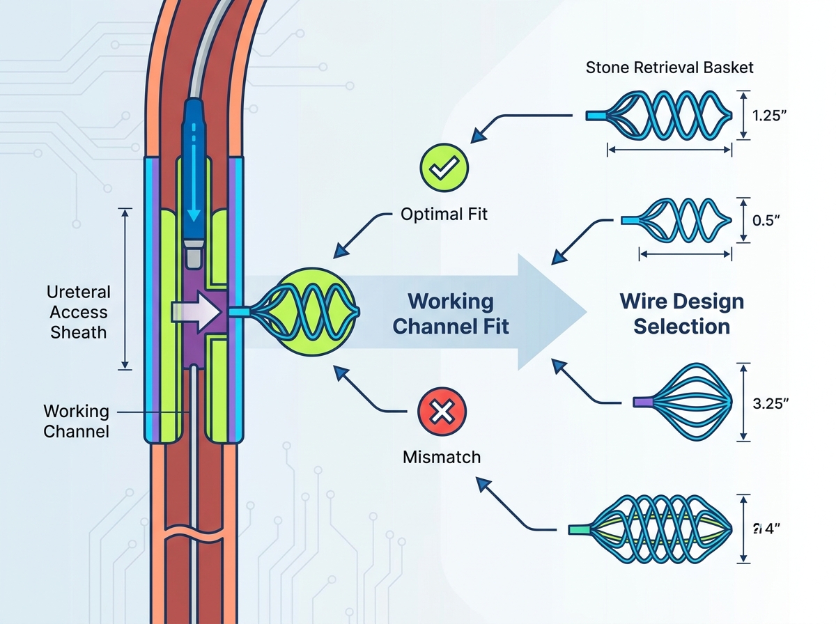
Instrument sizing in urology relies on the French (Fr) scale, where 1 Fr equals exactly 0.33 millimeters in diameter. The external diameter of the delivery sheath determines its compatibility with the endoscope's working channel. Standard sizing tiers generally fall into two categories: ultra-slim (1.3F to 1.5F) and standard (1.9F to 2.2F). In contemporary flexible ureteroscopy, 1.5F and 1.9F have emerged as the primary benchmarks. The choice between these sizes dictates the physical constraints of the procedure.
The "occupancy" calculation provides a mathematical framework for understanding these constraints. When an instrument is inserted into a working channel, it occupies a specific cross-sectional area, restricting fluid flow.
Working Channel Size | Basket Sheath Size | Occupied Area (%) | Impact on Irrigation Flow | Impact on Scope Deflection |
|---|---|---|---|---|
3.6 Fr | 1.5 Fr | ~17% | Minimal reduction; clear visibility maintained. | Negligible; full articulation preserved. |
3.6 Fr | 1.9 Fr | ~28% | Moderate reduction; adequate for routine clearance. | Slight resistance; minor loss of acute angles. |
3.6 Fr | 2.2 Fr | ~37% | Severe reduction; poor visibility during active bleeding. | High resistance; significant loss of lower pole access. |
This mathematical fit correlates directly to real-world clinical visibility. According to Poiseuille's law, flow rate is proportional to the fourth power of the radius. Therefore, even a fractional increase in sheath diameter causes a massive drop in irrigation flow. A smaller device, such as a 1.5F configuration, is mandatory when high-flow irrigation is required to clear dense stone dust or sudden bleeding. The ultra-slim profile maximizes the remaining open area within the channel, ensuring the surgeon maintains a clear visual field during extraction maneuvers.
System-level compatibility extends beyond the endoscope; it must include the entire access conduit. Proper ureteral access sheath selection directly impacts the efficiency of the extraction process. The inner diameter of the access sheath dictates the maximum size of the stone fragment that can be safely removed intact. If a captured stone exceeds the inner diameter of the access sheath, the surgeon cannot withdraw the loaded instrument without risking impaction or ureteral avulsion.
Multi-pass dynamics heavily rely on the friction coefficients between the interacting surfaces. During repeated extractions, the instrument sheath rubs against the working channel of the ureteroscope. Simultaneously, the external shaft of the ureteroscope rubs against the inner lumen of the access sheath. Hydrophilic coatings on these surfaces reduce friction, enabling smooth, rapid passes.
Intrarenal pressure considerations are paramount. The combined footprint of the scope and the extraction device within the access sheath affects fluid outflow. If the access sheath is completely occluded by the instruments, irrigation fluid cannot escape. This leads to dangerously elevated renal pelvic pressures. Selecting an appropriately sized access sheath ensures adequate outflow around the scope, mitigating the risk of pyelovenous backflow and postoperative sepsis.
The proximal handle serves as the surgeon's primary interface with the extraction device. Workflow optimization in modern urology frequently relies on the clinical utility of detachable handles. A detachable mechanism allows the surgeon to completely remove the handle assembly while leaving the wire engaged with a stone or foreign body.
This feature is essential when a captured stone proves too large to extract. By detaching the handle, the surgeon can slide the flexible ureteroscope off the instrument shaft, leaving the device in place as a safety wire. The surgeon can then introduce a rigid scope or a laser fiber alongside the shaft to fragment the entrapped stone. This capability transforms a potential surgical emergency into a manageable intracorporeal lithotripsy scenario.
Ergonomics and tactile feedback are equally important. The handle design must provide the surgeon with a 1:1 tactile response, ensuring precise control over the opening and closing tension of the cage. When a stone is engaged, the surgeon must feel the resistance through the handle to avoid applying excessive force that could sever the wire or crush a fragile calculus.
Standardization across fleets offers significant procurement and operational benefits. Standardizing on a single handle mechanism across different sizes and wire configurations reduces the learning curve for operating room staff. Consistent handle mechanics ensure that nurses and technicians can anticipate deployment behaviors, streamlining the surgical workflow and reducing the likelihood of intraoperative errors.
Basket entrapment represents one of the most severe complications in endoscopic stone management. The entrapment scenario typically occurs when a surgeon captures a stone that is larger than the ureteral orifice, the strictured ureteral lumen, or the inner diameter of the access sheath. Once engaged, the rigid calculus prevents the cage from collapsing back into its delivery sheath. If the entire assembly cannot be safely withdrawn, the device becomes anchored within the urinary tract.
Mitigation begins with intelligent design selection. Specific configurations, particularly tipless and highly flexible nitinol variants, allow for easier stone release. If extraction is not anatomically feasible, the surgeon can advance the sheath slightly to loosen the wires and manipulate the handle to release the stone back into the renal pelvis. Round wires, lacking the aggressive grip of flat wires, facilitate this release maneuver more readily.
When release is impossible, strict emergency protocols must be initiated. The primary technical step for managing an entrapped device involves utilizing holmium or thulium lasers. The surgeon must carefully advance a laser fiber alongside the entrapped wires and systematically fragment the calculus from within the cage. The detachable handle feature is vital here, allowing the removal of the primary scope to make room for dual-channel instruments or parallel access techniques. Extreme caution is required to avoid firing the laser directly onto the nitinol wires, which could sever the cage and leave metallic fragments within the kidney.
The lower pole of the kidney presents unique anatomical challenges. Retrieving stones from this region requires navigating acute infundibulopelvic angles that severely restrict instrument passage. When a flexible ureteroscope is fully deflected to reach a lower pole calyx, the working channel is bent at an extreme angle. Attempting to advance a stiff, large-diameter instrument through this tortuous path can cause mechanical failure of the endoscope's internal lining or snap the deflection pull-wires.
Mitigation via sizing is the most effective strategy for lower pole access. Surgeons must utilize ultra-slim (1.5F), highly flexible nitinol devices when targeting dependent calculi. The 1.5F profile easily navigates the acute bends of the deflected working channel without adding rigid resistance. This ensures that the scope maintains its maximum articulation, allowing the surgeon to reach the base of the calyx. Attempting to force a standard 1.9F or 2.2F flat-wire device into a fully deflected scope will almost certainly result in loss of access and potential damage to the capital equipment.
To safely navigate the lower pole, follow these procedural steps:
Advance the flexible ureteroscope into the renal pelvis before deflecting the tip.
Identify the target calyx and assess the infundibulopelvic angle.
Pre-load the 1.5F extraction device into the working channel, keeping the tip just inside the scope.
Deflect the scope to enter the lower pole calyx.
Deploy the cage slowly, utilizing the tipless design to sweep the mucosal floor.
No single extraction device is universally optimal for all clinical scenarios. Selection must be dictated by the specific stone burden, the anatomical location of the calculus, and the exact dimensional specifications of the endoscope fleet. Balancing fluid dynamics, scope deflection, and capture mechanics requires a deliberate, evidence-based approach to instrument procurement.
Procurement and clinical teams should prioritize 1.5F to 1.9F tipless nitinol configurations for routine flexible ureteroscopy. These devices offer the best balance of atraumatic manipulation, high first-pass capture rates, and preservation of working-channel flow. Stiffer, flat-wire designs should be reserved for semi-rigid ureteroscopy or specific sweeping maneuvers in the linear sections of the ureter. Reviewing in vitro retrieval capability data alongside real-world clinical outcomes will ensure that the selected devices meet the rigorous demands of modern endourology.
To optimize procedural success, take the following actions:
Audit your current endoscope working-channel dimensions to establish baseline compatibility metrics.
Review existing access sheath inventory to ensure inner diameters align with the maximum capture volumes of the selected extraction devices.
Request clinical evaluation samples of both round-wire and flat-wire nitinol configurations to assess tactile feedback and deployment mechanics in vivo.
Standardize handle mechanisms across the department to streamline staff training and reduce intraoperative deployment errors.
A: A tipless design eliminates the protruding distal wire. This allows the cage to open completely flush against the renal papilla. Surgeons can capture mucosal-embedded stones at the base of a calyx without causing urothelial trauma. It also improves overall scope deflection in tight anatomical spaces.
A: Larger French sizes increase the physical stiffness of the instrument. When inserted into the working channel, this stiffness resists the internal pull-wires of the flexible ureteroscope. This directly reduces the maximum deflection angle, making it difficult to access lower pole calculi.
A: Nitinol possesses unique shape-memory and superelastic properties. It resists permanent kinking, maintains perfect cage symmetry after multiple deployments, and withstands the mechanical stress of tortuous anatomy. Stainless steel is prone to plastic deformation and tangling during complex procedures.
A: The access sheath dictates the maximum size of fragments that can be safely extracted from the urinary tract. It also reduces friction during rapid, repeated passes and ensures adequate fluid outflow to maintain safe intrarenal pressures during the procedure.
A: Flat wires provide high radial dilation and aggressive sweeping power, making them ideal for heavy or impacted stones. Round wires offer gradual, atraumatic opening mechanics that conform to irregular shapes, minimizing tissue trauma during delicate calyceal manipulation.
A: Standard single-channel ureteroscopes lack the spatial capacity to accommodate both simultaneously. Concurrent use requires specialized dual-channel scopes or techniques where the handle is detached, allowing parallel access of a laser fiber alongside the deployed wire.
A: The initial protocol involves attempting to gently release the stone by advancing the sheath. If unsuccessful, the surgeon can disassemble the detachable handle, remove the scope, and utilize intracorporeal holmium laser lithotripsy to fragment the stone within the wires.
